

BREAKING NEWS
Site Under Development, Content Population and SEO, Soft Launch 1st January 2020
Penile Intraepithelial Neoplasia (PeIN or PIN), also known as penile squamous cell carcinoma, is a premalignant condition of the penile epithelium. The most sensitive way of detecting PeIN is by applying acetic acid to the skin of the male genitalia. The treatment procedures for PeIN include topical chemotherapy, laser therapies (Nd:YAG, and Co2), cryotherapy, photodynamic therapy, and surgical excision.
Credit: Evgeniy Kalinovskiy/ Shutterstock.com
Topical therapy is used to treat penile lesions such as Pseudoepitheliomatous, Keratotic and Micaceous Balanitis, Bowenoid papulosis (BP), and Carcinoma in situ (CIS).
During the period of treatment, the affected part becomes wadded and inflamed. Topical corticosteroids can be used additionally for the discomfort; however, reactive changes in certain regions can take four to eight weeks to heal. The number of patients cured by undergoing this type of treatment is minimal (less than ten patients).
In laser therapy, neodymium-doped yttrium aluminium garnet (Nd:YAG) and carbon-dioxide (CO2) lasers are used. This is the first-line treatment procedure that is predominantly involved in treating BP and CIS. Treatment involving these lasers has minor complications such as bleeding with a little pain at the laser ablated sites.
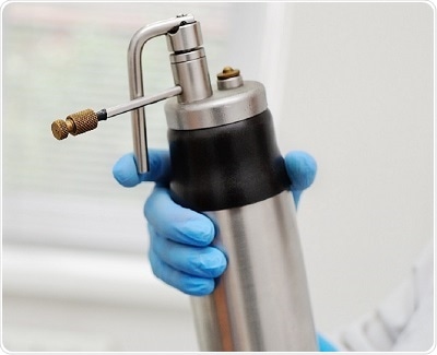
Cryotherapy utilizes nitrous oxide or liquid nitrogen to cause tissue damage at temperatures between 20 and 50°C. Tissue damage is achieved by the development of ice crystals resulting in cell disruption and cell death. This treatment of penile lesions has a higher risk of recurrence compared with surgical excision and chemotherapy procedures.
Photodynamic therapy (PDT) uses a photosensitizing cream with a chemical called delta-5-aminolaevulinic acid, applied in the affected area for nearly three hours. Following the exposure of PDT light to lesions, cell death of excited cells occurs. This therapy is performed for a period of approximately 35 months.
Surgical excision is the ideal procedure to treat all premalignant lesions. This procedure is recommended in patients who are unwilling to follow instructions and strict treatment protocols.
Circumcision plays a crucial role in the management of precancerous conditions. It helps in complete removal of lesions present on the foreskin and avoids HPV infection, advancement to invasive disease, and chronic inflammation. A 5% of acetic acid is smeared on the penis for about 5 min to identify the occult regions of CIS with the help of acetowhite reaction and malignant resection.
A split-thickness skin graft taken from the thigh is utilized to protect the exposed glans. This method provides conservation of the penile form, length, and function. It also has good oncological regulation with better cosmetic appearance.
Another surgical procedure used for excision is Mohs micrographic surgery. This surgery helps in complete removal of a lesion in thin sections with high preservation of penile tissue. Since it is a time-consuming and difficult process associated with a recurrence rate of 32%, it is not a widely used technique.